Saturday, November 14, 2009

Scientific Paper Review

Vocal Fold Nodules: Morphological and Immunohistochemical Investigations

Authors: Regina Helena Garcia Martins, Julio Defaveri, Maria Aparecida Custo´ dio Domingues,
Rafael de Albuquerque e Silva, and Alexandre Fabro.

*View the article by clicking here.*

Introduction

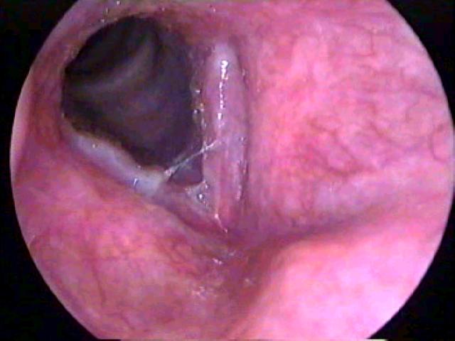
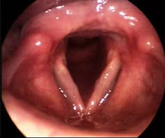
Vocal fold nodules are bilateral epithelial thickenings located in the mid third of the vocal folds. They occur after traumatic and constant collision of the delicate vocal mucosa. These lesions are a common cause of hoarseness, and occur frequently in people who overuse their voices or use them professionally, such as children, teachers, singers and speakers. They may also be induced by a variety of other pre-disposing factors. Vocal fold nodules are often confused in histopathological examinations with other benign laryngeal lesions.

The purpose of this study was to examine the morphological and immunohistochemical characteristics of vocal fold nodules. A combined assessment using immunohistochemistry, light microscopy and electron microscopy was employed to reveal morphological details essential for characterization of vocal fold nodules.


Histological Study


Light Microscopy

Light microscopy was used to review 15 histological slides stained with hematoxylin and eosin. These slides were created from biopsies provided by patients submitted to surgery in the Voice Disorders Ward of the Medical School of Botucatu, Brazil from 2001 to 2006.

The epithelium and lamina propria were examined, and a semiquantitative score of 0 (no alteration), 1 (mild alteration), 2 (moderate alteration), and 3 (intense alteration) was used to qualify inspection of the slides.

Light microscopy studies of these vocal fold nodules recorded a predominance of epithelial hyperplasia (an abnormal increase in the number of cells, and consequential enlargement), basement membrane thickening, fibrosis (formation of excessive fibrous connective tissue), and lamina propria edema (swelling).


Electron Microscopy

Transmission and scanning electron microscopy (TEM, SEM) studies included five new prospective surgical cases of vocal fold nodules. Of each, one nodule was sent for SEM, and the contralateral nodule was sent for TEM analysis. Tissue preparation was performed according to conventional methods.

Ultrastructural SEM analysis showed a reduction in mucous lacing and an increase in scaled cells. TEM analysis showed hyperplasia of the epithelium, enlargement of the intercellular junctions, which were filled with fluid, and weakness in the basal membrane. All specimens showed subepithelial thickening of the lamina reticularis, distorting the basal cell layer.

Basically, through SEM and TEM ultrastructural analyses, an enlargement in the cell junctions, alterations in desmosome structure, basement membrane break points, and thickening of the lamina reticularis was noticed.


Immunohistochemistry

Immunohistochemical evaluation was performed on the 15 surgical vocal nodule slides previously examined by light microscopy. Three slides of normal vocal fold tissue recovered during the autopsy of three patients were used as the experimental control.

Slides were treated with antifibronectin polyclonal antibody, antilaminin monoclonal antibody, and anticollagen IV monoclonal antibody using the Avidin Biotin Peroxidase method. They were then examined by an experienced pathologist at different magnifications. Results were interpreted, depending on the degree of color intensity, as mild, moderate, or intense.

This immunohistochemical analysis, revealed a marked immunoexpression of fibronectin on the basement membrane and on the lamina propria.


Discussion

This research highlighted a number of morphological characteristics of vocal fold nodules. This is important, as the pathophysiology of these laryngeal lesions is still little understood.

Significant findings of this histological study include epithelial layer thickening and the increase in fibronection in the basement membrane in vocal nodules. In the vocal folds, the basement membrane is adhered to the basal cell plasmatic membrane by anchoring fibers. These fibers connect the lamina densa to the superficial layers of the lamina propria. Alterations of the anchor fibers result in basement membrane alteration, resulting in the opening of spaces, which are later filled with a heterogeneous material. This likely represents a response to phonatory trauma.

The SEM study showed a higher number of scaling cells, also probably resulting from excessive collision during vocalization. The subepithelial edema noticed through light microscopy was also seen by scanning microscopy, and was represented by a reduction in the mucosal lace-like pattern because of epithelial expansion. Many scientific papers have noted the characteristics of a high prevalence of subepithelial edema, loss of intercellular junctions, and epithelial dysplasia in other types of benign laryngeal lesions, such as Reinke’s edema. Therefore, these histological alterations are not specific to vocal fold nodules.


Critique

This paper is useful in that it confirms some key morphological characteristic of vocal fold nodules. Unfortunately, the histological study did not reveal any new information about the lesions, stopping just short of discovering some very interesting biochemical information.

Additional immuno-electron microscopic evaluation would have been useful in clarifying the real composition of the supepithelial heterogenous deposits (which resulted in the thickening of the lamina reticularis) seen consistently in all TEM specimens. Had this been performed, novel information would have been revealed and the whole paper would have been much more striking.

The light microscopy study was performed predominantly on old slides, stained between 2001 and 2006. The paper was published in 2009, which means that some of the slides examined were up to 8 years old. The conditions in which they were stored for 8 years were not specified. The use of such old specimens is understandable considering how difficult it is to get a surgical vocal fold nodule tissue samples, as these lesions are principally treated with non-invasive methods. However, this experiment could have been improved by using fresh samples that were newly stained. This would ensure that the observations made were not artifacts, results of long-time neglect. Otherwise, information about the storage conditions of the slides should have been included.

The statistical methods, and laboratory techniques used in this evaluation were all sound, and overall, the research was well thought out and organized. Unfortunately, for Martins et al., it revealed no new information about vocal fold nodules, and the pathophysiology of these benign lesions remains little understood.


Reference

Martins, R. H. G, Defaveri, J., Custo´ dio Domingues, M. A., Albuquerque e Silva, R. de, & Fabro, A. (January 2009). Vocal Fold Nodules: Morphological and Immunohistochemical Investigations. Journal of Voice. doi:10.1016/j.jvoice.2009.01.002

Disclaimer: All figures displayed in this blog post were borrowed from the scientific paper indicated above, and all rights are the property of the paper, not this website.

Tuesday, October 27, 2009

My Favorite Tissue: The Vocal Folds


The Role of Voice in Daily Life

Voice is our primary means of expression. Combined with facial expressions and hand gestures, it signals who we are, what we want and how we feel. There is no critical age for vocalization; it begins from the moment of birth. As we grow our voices change in accordance to reflect our culture, personalities, conditions of health, age and gender.

Vocal expression is used primitively as a means for survival. Expressions of pain and hunger are often vocal, especially in infants. Offensively, voice is used to startle and threaten. The roar of a lion, trumpet of an elephant are quite intimidating. Vocalization is also used as a means to locate someone or to express emotion. The six primary emotions- fear, anger, joy, sadness, surprise, and disgust- are all expressible vocally.

The role of voice in speech is obvious. Personality is reflected in voice, but especially in speech, the use of voice with language. Confidence and authority can be judged by rate of speech, number of pauses, and volume. Slow speech can be considered thoughtful, in the positive sense, or a mark of incompetence.

The voice also offers a window into health and fitness of an individual. Hoarseness is often a sign of viral infection. Vocal tremors can be an early sign of a neurological disease. Change in pitch could indicate a fluid imbalance. A ‘strangled’ voice is a sign of severe emotional trauma.

The vocal folds are the epicenters of sound production. The vibration of this tissue is responsible for phonation, and an awareness of its histology and pathology is essential for our understanding of voice and communication.


Basic Anatomy of the Larynx

The larynx is an organ in the center of a highway of supply lines. It is a musculo-cartilaginous group situated in the neck, in front of the spinal cord, where blood vessels, nerves, glands, and other critical supply lines of the human body surround it.

The larynx sits as an oddly shaped box atop the cricoid cartilage of the trachea. The cricoid cartilage forms a complete, strong ring and articulates with the thyroid and arytenoid cartilages and attaches below to the first cartilage of the trachea.


The ability of the laryngeal cartilages to articulate is essential for the larynx as a valve and organ of phonation. This is accentuated by the position of the vocal cords housed at its centre. The vocal chords stretch from the arytenoid cartilage posteriorly, to the inner surface of the thyroid cartilage in front. The triangular shape of the glottic airway occurs because the vocal cords join in front (the anterior commissure), but are able to part in the back (the posterior commissure).

The suspension of the larynx in the neck involves a total of ten muscle groups (the specifics of which go beyond the scope of this assignment). The amazing range of motion that they allow can be felt by placing a finger along the midline of your neck during swallowing, breathing and phonation.

The interior of the larynx is exposed to all airway fluids swept upwards by ciliary movement from the lungs. The larynx itself is lined by ciliated mucous secreting epithelium. Mucous glands occur especially heavily in the ventricles between the true and false vocal cords to keep them moist and lubricated. Below is a cross section of the larynx. Note the round vestibular spaces between the false vocal cords (above the space) and the true vocal cords (below the space).
Fun Fact!
The speech skill is a wonder. To produce a phrase, about 100 muscles of the chest, neck, jaw, tongue and lips must collaborate.


The Histological Composition of the Vocal Folds

A vertical, frontal section of the larynx in the following figure reveals the two vocal folds (9, 11, 12, 13), their supporting cartilage (4, 6) and muscle (10).

The laryngeal ventricle (10) is a deep indent that separates the false vocal fold (9) from the true vocal fold (11, 12, 13). The lumen is lined with pseudostratified columnar epithelium with goblet cells.

The mucosa in the wall of the ventricle (3, 8) is similar to that of the false vocal fold (9). In the lamina propria (3) are mixed glands (8) that are predominantly mucous. Lymphatic nodules (2) are also located in the lamina propria on the ventricular side of the vocal fold and are sometimes called the laryngeal tonsils. The lamina propria (3) blends with the perichondrium (5) of the hyaline thyroid cartilage (4), and no distinct submucosa is found. The lower wall of the ventricle transitions into the true vocal fold (11, 12, 13).

The mucosa of the true vocal fold (11, 12, 13) is composed of five layers of tissue, as seen in following figure. The most superficial layer is nonkeratinized stratified squamous epithelium. This layer is extremely thin (0.1mm thick), and gives the vocal folds the glistening white appearance seen during laryngoscopic examination.

Deep to this layer is the lamina propria, which is composed of three distinct tissues, as seen in the figure to the right. The first layer of the lamina propria (the second layer of vocal fold tissue) is known as the superficial layer. This layer is approximately 0.5 mm thick and is made up of elastin fibers, which permit it to stretch extensively. The intermediate layer of lamina propria is 1-2mm thick. It is also composed of elastin fibers, but these fibers have a more or less anterior-posterior orientation as opposed to the more or less random orientation found in the superficial layer. The final, deep layer of lamina propria is also 1-2mm thick and is composed of collagen fibers, which prohibit extension.

Deep to the deep lamina propria is the fifth layer of the vocal folds, the skeletal thyrovocalis muscle (often abbreviated vocalis). This muscle makes up the bulk of the vocal fold, and is oriented in an anterior-posterior course. The vocalis muscle is controlled voluntarily and is innervated by a branch of the X Vagus nerve. The vocalis is antagonistic to the cricothyroid muscle. Contraction of the vocalis will draw the thyroid and cricoid cartilages farther apart in front, while contraction of the cricothyroid muscle draws them closer together. This allows the vocal cords to be tensed for sound production.

Refering back to the first histological figure of this section, the vocal ligament (12) is found at the apex of the true vocal fold. It is composed of dense elastic fibers. From that point downwards, the mucosa returns to pseudostratified columnar epithelia with mixed glands (14, 15). The hyaline cricoid cartilage is the lowermost catilage of the larynx (6).

Fun Fact!
The mass of the vocal cord is about one gram.

Fun Video!
Watch this laryngoscopy on YouTube before you read the next section.


Vocal Fold Oscillation

The biomechanics of vocal tissue oscillation to produce sound is very complex. In spoken communication, a steady flow of air from the lungs is used to generate acoustic power. Airflow is converted into oscillatory motion in the larynx at the vocal folds. They are set into vibration, like flags in the wind, by bringing them into and out of the airstream.

This invokes the Bernoulli effect, a principle of physics that states that given a constant volume of airflow, at a point of constriction there will be a decrease in air pressure perpendicular to the flow, and an increase in velocity of the flow. Airflow increase and air pressure drop cause the vocal folds to undergo cycles of vibration, as seen in the figure below.

Basically, the motion of the tissue and the airflow disturb the molecules of air and produce the phenomenon we call sound. The sound then propagates along the airstream towards the mouth and radiates into space. Without the vocal folds, air would move relatively unimpeded out of the lungs and into the oral cavity.

The mathematical and physical theories explaining voice and song are a very complex inter-disciplinary field of science. Please see the text Principles of Voice Production for a full and detailed account of these ideas if you are interested.

Fun Fact!
No other animals can copy the sounds of our own speech and understand its meaning - not even our nearest relatives, the chimpanzees.


Common Vocal Fold Pathologies

The voice acts as a window into health and fitness of an individual. The unusual pitch or tone of a patients voice is often used as a symptom for the diagnosis of whole body health issues like viral infections, fluid imbalances, neurologic disease and emotional trauma. Some common vocal fold pathologies include the following:

Congenital Voice Disorders

These are structural abnormalities in the airway system. A common example is the laryngeal web. This occurs when the vocal folds fail to completely separate during fetal development. Children with this abnormality experience difficulty breathing and an unusual cry. The web must be removed surgically and a stent placed between the folds until the epithelium has healed over completely.

Tissue Infection

A very common voice disorder is acute laryngitis. It is frequently associated with a cold or influenza, and is the result of a viral infection. The initial immune response of increased blood flow to the area changes the surface of the vocal folds, making them become red and thickened. Further into the healing process white blood cells remove debris from the tissue. This makes the vocal folds more viscous and difficult to move, resulting in the characteristic rough voice.Vocal Nodules

A vocal fold nodule is a mass of callous-like tissue that grows on the vocal folds. The formation of nodules on the vocal folds is a reaction to the localized mechanical stress of collision between the folds. Abuse is generally caused by excessive screaming, prolonged talking, or chronic faulty singing technique. Masses will typically appear where contact is most forceful.

Symptoms include painful speech production, hoarseness of speech, frequent voice cracks and a decreased vocal range. A nodule reduces or obstructs the ability of the vocal folds to create the rapid changes in air pressure, which generate human speech. Nodules are generally bilateral, and are initially soft, and easily treatable with voice therapy. They can however, become hardened and fibrous with prolonged misuse, at which point surgery is required to regain normal vocalization.

Vocal Polyps

Very similar to vocal nodules, vocal polyps are also caused by misuse of the voice. They have the same symptoms and treatment, but occur as more of a blister-like growth on the vocal folds, rather than as callous-like growth as in the case of nodules.Contact Ulcers

Contact ulcers are unilateral or bilateral erosions of the mucous membrane over the vocal folds. They are caused by voice abuse in the form of repeated sharp glottal attacks and are often experienced by singers. They may also occur after endotracheal intubation if an oversized tube abrades the mucosa of the vocal process.

Vocal Paralysis

Vocal fold paralysis is a neurological disorder. It occurs when one or both of the vocal folds do not open or close properly. This disorder may be caused by head trauma, a neurologic disorder, a neck injury, cancer, a tumor pressing on a nerve, or a viral infection, though in many cases the cause is unknown.

If unilateral paralysis occurs, the voice is usually hoarse or breathy. With one vocal cord functioning normally, it is possible to increase its amplitude of vibration and meet the paralyzed fold at the midline. Bilateral paralysis of the vocal cords, although rare, usually causes people to have difficulty breathing as paralysis impairs the ability to protect the airway, and breath properly.


Surface Irritation

The leading example of vocal fold surface irritation is smoking. Repeated irritation of the epithelial surface of the vocal folds with smoke can cause chronic inflammation leading to altered tissue shape, elasticity and viscosity. Gastroesophageal reflux may also cause irritation of the vocal fold tissue as stomach acid spills into the larynx. Symptoms include hoarseness, and treatment is removal of the irritant. Persistent irritation can lead to carcinoma of the larynx.

Vocal Fold Cancer

Like other cancers, cancer of the vocal folds is caused by normal tissue changing into tissue that grows uncontrollably. If unchecked, vocal fold cancer can spread into deeper tissues, to the neck outside of the larynx and eventually to other parts of the body, principally the lungs. Smoking is responsible for the overwhelming majority of laryngeal cancers. Laryngeal cancer in nonsmokers is a rarity.

References

Eroschenko, V. P. (2005). diFiore’s Atlas of Histology with Functional Correlations (10th ed.). Baltimore: Lippincott Williams & Wilkins.

Proctor, D. F. (1980). Breathing, Speech and Song. New York: Springer- Verlag/ Wein.

Seikel, J. A., King, D. W., & Drumright, D. G. (2000). Anatomy and Physiology for Speech, Language, and Hearing (2nd ed.). San Diego: Singular Publishing Group Inc.

Titze, I. R. (1994). Principles of Voice Production. New Jersey: Prentice Hall.

Disclaimer All images from this blog are linked to their internet source, and all rights to these images belong to the source websites, not this one.