Authors: Regina Helena Garcia Martins, Julio Defaveri, Maria Aparecida Custo´ dio Domingues,
Rafael de Albuquerque e Silva, and Alexandre Fabro.
*View the article by clicking here.*
Introduction
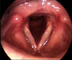
Vocal fold nodules are bilateral epithelial thickenings located in the mid third of the vocal folds. They occur after traumatic and constant collision of the delicate vocal mucosa. These lesions are a common cause of hoarseness, and occur frequently in people who overuse their voices or use them professionally, such as children, teachers, singers and speakers. They may also be induced by a variety of other pre-disposing factors. Vocal fold nodules are often confused in histopathological examinations with other benign laryngeal lesions.
The purpose of this study was to examine the morphological and immunohistochemical characteristics of vocal fold nodules. A combined assessment using immunohistochemistry, light microscopy and electron microscopy was employed to reveal morphological details essential for characterization of vocal fold nodules.
Histological Study
Light Microscopy
Light microscopy was used to review 15 histological slides stained with hematoxylin and eosin. These slides were created from biopsies provided by patients submitted to surgery in the Voice Disorders Ward of the Medical School of Botucatu, Brazil from 2001 to 2006.
The epithelium and lamina propria were examined, and a semiquantitative score of 0 (no alteration), 1 (mild alteration), 2 (moderate alteration), and 3 (intense alteration) was used to qualify inspection of the slides.
Light microscopy studies of these vocal fold nodules recorded a predominance of epithelial hyperplasia (an abnormal increase in the number of cells, and consequential enlargement), basement membrane thickening, fibrosis (formation of excessive fibrous connective tissue), and lamina propria edema (swelling).

Electron Microscopy
Transmission and scanning electron microscopy (TEM, SEM) studies included five new prospective surgical cases of vocal fold nodules. Of each, one nodule was sent for SEM, and the contralateral nodule was sent for TEM analysis. Tissue preparation was performed according to conventional methods.
Ultrastructural SEM analysis showed a reduction in mucous lacing and an increase in scaled cells. TEM analysis showed hyperplasia of the epithelium, enlargement of the intercellular junctions, which were filled with fluid, and weakness in the basal membrane. All specimens showed subepithelial thickening of the lamina reticularis, distorting the basal cell layer.
Basically, through SEM and TEM ultrastructural analyses, an enlargement in the cell junctions, alterations in desmosome structure, basement membrane break points, and thickening of the lamina reticularis was noticed.


Immunohistochemistry
Immunohistochemical evaluation was performed on the 15 surgical vocal nodule slides previously examined by light microscopy. Three slides of normal vocal fold tissue recovered during the autopsy of three patients were used as the experimental control.
Slides were treated with antifibronectin polyclonal antibody, antilaminin monoclonal antibody, and anticollagen IV monoclonal antibody using the Avidin Biotin Peroxidase method. They were then examined by an experienced pathologist at different magnifications. Results were interpreted, depending on the degree of color intensity, as mild, moderate, or intense.
This immunohistochemical analysis, revealed a marked immunoexpression of fibronectin on the basement membrane and on the lamina propria.
Discussion
This research highlighted a number of morphological characteristics of vocal fold nodules. This is important, as the pathophysiology of these laryngeal lesions is still little understood.
Significant findings of this histological study include epithelial layer thickening and the increase in fibronection in the basement membrane in vocal nodules. In the vocal folds, the basement membrane is adhered to the basal cell plasmatic membrane by anchoring fibers. These fibers connect the lamina densa to the superficial layers of the lamina propria. Alterations of the anchor fibers result in basement membrane alteration, resulting in the opening of spaces, which are later filled with a heterogeneous material. This likely represents a response to phonatory trauma.
The SEM study showed a higher number of scaling cells, also probably resulting from excessive collision during vocalization. The subepithelial edema noticed through light microscopy was also seen by scanning microscopy, and was represented by a reduction in the mucosal lace-like pattern because of epithelial expansion. Many scientific papers have noted the characteristics of a high prevalence of subepithelial edema, loss of intercellular junctions, and epithelial dysplasia in other types of benign laryngeal lesions, such as Reinke’s edema. Therefore, these histological alterations are not specific to vocal fold nodules.
Critique
This paper is useful in that it confirms some key morphological characteristic of vocal fold nodules. Unfortunately, the histological study did not reveal any new information about the lesions, stopping just short of discovering some very interesting biochemical information.
Additional immuno-electron microscopic evaluation would have been useful in clarifying the real composition of the supepithelial heterogenous deposits (which resulted in the thickening of the lamina reticularis) seen consistently in all TEM specimens. Had this been performed, novel information would have been revealed and the whole paper would have been much more striking.
The light microscopy study was performed predominantly on old slides, stained between 2001 and 2006. The paper was published in 2009, which means that some of the slides examined were up to 8 years old. The conditions in which they were stored for 8 years were not specified. The use of such old specimens is understandable considering how difficult it is to get a surgical vocal fold nodule tissue samples, as these lesions are principally treated with non-invasive methods. However, this experiment could have been improved by using fresh samples that were newly stained. This would ensure that the observations made were not artifacts, results of long-time neglect. Otherwise, information about the storage conditions of the slides should have been included.
The statistical methods, and laboratory techniques used in this evaluation were all sound, and overall, the research was well thought out and organized. Unfortunately, for Martins et al., it revealed no new information about vocal fold nodules, and the pathophysiology of these benign lesions remains little understood.
Reference
Martins, R. H. G, Defaveri, J., Custo´ dio Domingues, M. A., Albuquerque e Silva, R. de, & Fabro, A. (January 2009). Vocal Fold Nodules: Morphological and Immunohistochemical Investigations. Journal of Voice. doi:10.1016/j.jvoice.2009.01.002
Disclaimer: All figures displayed in this blog post were borrowed from the scientific paper indicated above, and all rights are the property of the paper, not this website.
